Open the structure
Download a PDB or mmCIF file from a structure database and drop it here, or open the crambin sample.
Open protein and nucleic acid structures from PDB or mmCIF files and explore them as cartoons, sticks or surfaces.
Find residues in the sequence, show ligands and measure distances, angles and dihedrals.
Loading the structure editor…
Download a PDB or mmCIF file from a structure database and drop it here, or open the crambin sample.
Switch between cartoon, ball and stick and space-filling, add a surface, and colour by chain, secondary structure, residue or B-factor.
Click residues in the sequence to zoom to them, label them, and measure distances, angles and dihedrals between atoms.
Turn molecules into illustrations, cover art and explainer clips for slides, papers and lessons with WaveSpeed AI.
Create journal-cover style art and lab illustrations from a description.
Animate an illustration into a short explainer clip.
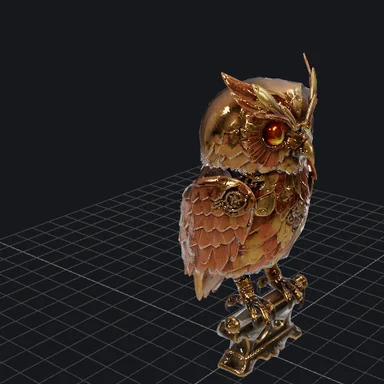
Make a 3D model of lab equipment or a stylised molecule.
$1 in free credits for eligible new accounts. No card needed. Generators have separate account and credit requirements. The structure drawer is free.
Yes, with no account and no limits beyond your device's memory.
No. Download the file from a structure database yourself and open it here; the viewer reads it in your browser and never uploads it.
Typical proteins and complexes open in a second or two. Very large assemblies with hundreds of thousands of atoms work on desktops but can be slow on phones; turn the surface off for those.
Ligands are drawn as ball and stick on top of the cartoon and listed in the panel; waters are hidden from the ligand list and the sequence.
PDB and mmCIF · Sequence and ligands · Files stay on your device