Open your images
Drop .dcm files, a whole folder (a CD or USB export works) or a ZIP. Images are grouped by patient, study and series.
View CT, MR, X-ray and ultrasound images in your browser, from single files, whole folders or ZIP archives.
Window and level, measure in millimetres and Hounsfield units, reformat volumes, read every tag, and remove patient details before you share.
Not for diagnostic use
Single .dcm files, a whole study folder from a CD or USB export, or a ZIP archive. Everything is read in your browser.
Files without the .dcm extension work too. Other files in the folder are skipped.Drop .dcm files, a whole folder (a CD or USB export works) or a ZIP. Images are grouped by patient, study and series.
Scroll through slices, drag to adjust window/level, zoom and pan, and measure lengths, angles and regions. Switch to 2 or 4 viewports or reformat a CT or MR volume.
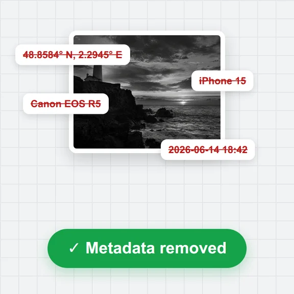
Download an anonymized copy with names, IDs and dates removed, or save images as JPG, PNG or an MP4 cine.
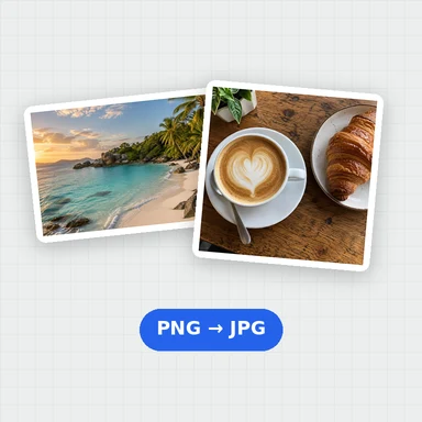
Turn findings into simple illustrations for patients, students or presentations with WaveSpeed AI.
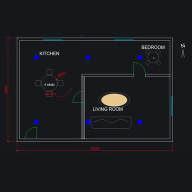
Create clear anatomy-style illustrations and diagrams for slides and handouts.
Turn an illustration into a short explainer clip.
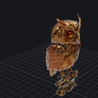
Make a 3D model to show shape and position.
$1 in free credits for eligible new accounts. No card needed. Generators have separate account and credit requirements. The DICOM viewer is free.
Yes. Open as many studies as you like, with no account, no watermark and no file limits beyond your device's memory.
No. This viewer is not a medical device and is not for diagnostic use. Use it to look at your own scans, to study, to teach or for research. Clinical reading must be done on certified software and displays.
No. Files are read and decoded in your browser and never leave your device. Some compressed formats need a decoder that downloads once from a public code library the first time it is used; your images are not sent anywhere.
DICOM files with or without the .dcm extension, folders from a CD, DVD or USB export (the DICOMDIR index is skipped and the images are read directly), and ZIP archives. Uncompressed, deflated, RLE, JPEG baseline and 12-bit, JPEG lossless, JPEG-LS, JPEG 2000 and HTJ2K images are supported, including multi-frame and colour images. Video-encoded DICOM (MPEG-2, H.264, HEVC) and JPEG XL are not.
Choose the ellipse or rectangle tool and drag over the area. The mean, standard deviation, minimum, maximum and area are shown in Hounsfield units on CT, using the rescale slope and intercept in the file. Lengths and areas use the pixel spacing; when an X-ray only has detector spacing, the viewer says so because magnification is not corrected.
It follows the DICOM PS3.15 Basic Application Level Confidentiality Profile: names, IDs, dates, institution and device details, free-text descriptions and private tags are removed or emptied, and every UID is replaced consistently across the whole export. You can choose to shift dates by a fixed number of days, keep patient age and sex, or keep series descriptions. Pixel data is copied unchanged.
No. Text burned into the pixels, common on ultrasound and screen captures, stays. The viewer warns you when a series is likely to have it; check those images before sharing.
Yes. For CT and MR volumes of parallel slices, choose MPR to see axial, coronal and sagittal views side by side with linked crosshairs. Click in any view to move the other two.
It doesn't do 3D volume rendering, PET/CT fusion, hanging protocols, structured reports, DICOM networking (PACS query or send) or writing to CD. It is for viewing, measuring, anonymizing and exporting.
Free, private and nothing to install.
/filters:quality(82)/media/images/1791123751656780930_IfFPY7R0.webp)
/filters:quality(82)/media/images/1791123737177812425_RjKT3cku.webp)
/filters:quality(82)/media/images/1791123748309870128_xlkNgK8N.webp)
/filters:quality(82)/media/images/1791123738630744263_oR09R0aj.webp)
/filters:quality(82)/media/images/1791177033162457776_uQ09OY7g.webp)
/filters:quality(82)/media/images/1791123734319125607_JdnwGPY7.webp)
/filters:quality(82)/media/images/1791123746355630206_udnxGPY7.webp)
/filters:quality(82)/media/images/1791123744771362580_yC0sPezZ.webp)
/filters:quality(82)/media/images/1791123738606970169_5KIR1ais.webp)
/filters:quality(82)/media/images/1791123748197655725_EUmuFNW6.webp)